


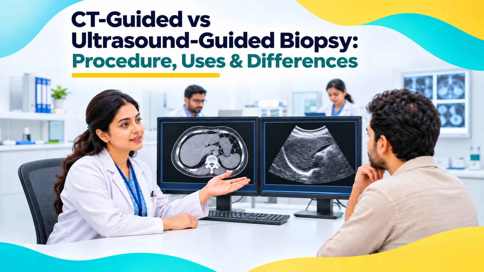
If a scan has found a lump or an unusual area, your doctor may recommend a biopsy to find out exactly what it is. Today, most biopsies are done with a needle, guided by imaging such as CT or ultrasound, rather than through surgery.
This guide explains what an image-guided biopsy is, how CT-guided and ultrasound-guided biopsies differ, when each is used, and what to expect before, during and after the procedure.
What Is an Image-Guided Biopsy?
An image-guided biopsy is a procedure in which a doctor uses imaging to guide a thin needle into a lump or organ and remove a small sample. The sample is then examined in a laboratory to make a diagnosis. These procedures are usually performed by a radiologist trained in interventional radiology.
Why Imaging Is Used
Imaging allows the doctor to:
- See exactly where the target is
- Plan a safe path that avoids blood vessels and other organs
- Confirm the needle is in the right place before taking a sample
This makes biopsies more accurate and safer, and avoids the need for surgery in most cases.
What Is a CT-Guided Biopsy?
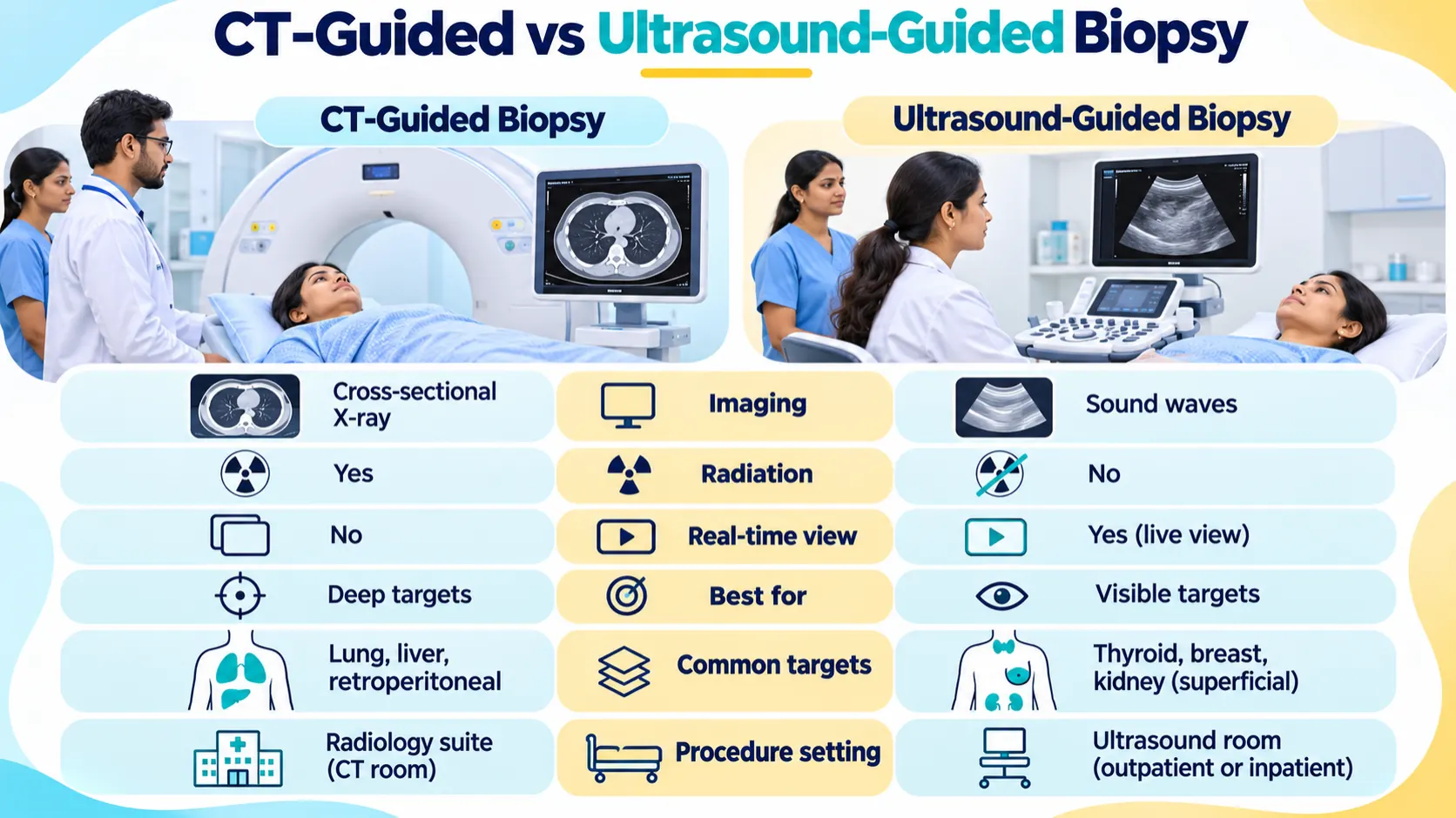
In a CT scan guided biopsy, a CT scanner takes cross-sectional images of the body while the needle is advanced in small steps, with images checked to confirm the needle's position. Some centres also use CT fluoroscopy, which gives real-time or near-real-time images during the procedure.
CT is useful for targets deep inside the body, or in areas that ultrasound cannot see clearly, such as the lungs, bones and areas behind bowel gas.
What Is an Ultrasound-Guided Biopsy?
In an ultrasound-guided biopsy, sound waves create live images on a screen. The doctor watches the needle in real time as it moves to the target.
Ultrasound is ideal for organs and lumps it can see clearly, such as the thyroid, breast, lymph nodes, liver, kidney and soft tissue lumps. It uses no radiation.
CT vs Ultrasound Comparison
When CT Guidance Is Preferred
- Lung lesions
- Bone lesions
- Deep abdominal or pelvic lesions
- Targets hidden behind bowel gas or bone
- Lesions that are not visible on ultrasound
When Ultrasound Guidance Is Preferred
- Thyroid nodules
- Breast lumps
- Enlarged lymph nodes in the neck, armpit or groin
- Liver and kidney lesions that can be seen on ultrasound
- Soft tissue lumps
- When avoiding radiation is important, such as in pregnancy or for young people, provided ultrasound can show the target and offers a safe route
FNAC vs Core-Needle Biopsy
CT versus ultrasound describes how the needle is guided. FNAC versus core biopsy describes the type of needle and sample:
- FNAC (fine needle aspiration cytology): a very thin needle collects cells. It is quick and gentle, but sometimes the sample is not enough for a full diagnosis.
- Core-needle biopsy: a slightly larger needle removes small cylinders of tissue. This shows how the cells are arranged, which helps with a more complete diagnosis, such as the type of a tumour.
Either type of needle can be used with CT or ultrasound guidance.
Preparing for an Image-Guided Biopsy
- You may need blood tests to check your blood clotting
- You may be asked not to eat for a few hours beforehand, depending on the biopsy site
- Tell your doctor about all your medicines, especially blood thinners
- Tell your doctor about allergies and whether you are or might be pregnant
- Arrange for someone to take you home if you will be given sedation
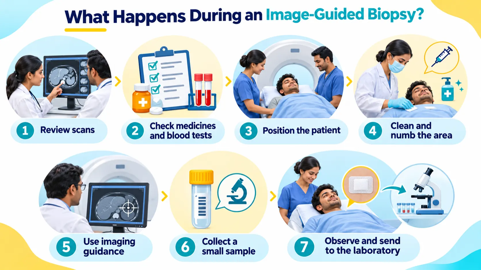
Procedure Step by Step
- You lie in a comfortable position for the target area.
- The doctor uses imaging to find the target and plan the safest path.
- The skin is cleaned, and a local anaesthetic is injected to numb the area.
- A small nick may be made in the skin.
- The needle is guided to the target using CT or ultrasound.
- One or more samples are taken.
- The needle is removed, pressure is applied, and a small dressing is placed.
Anaesthesia and Discomfort
Most biopsies are done under local anaesthesia, so you are awake. You may feel a sting from the numbing injection and some pressure when samples are taken. Sedation may be offered for some deeper biopsies.
Recovery and Receiving Results
You will rest for a short time afterwards so the team can check for bleeding or other problems. This may be longer after lung, liver or kidney biopsies, and a chest X-ray may be done after a lung biopsy. Most people go home the same day.
Results usually take several days, sometimes longer if special tests are needed. Your doctor, or a specialist such as an oncologist if needed, will explain the results and next steps.
Risks and Complications
Image-guided biopsies are generally safe. Possible risks include:
- Pain or bruising at the biopsy site
- Bleeding, usually minor
- Infection, which is rare
- A sample that is not enough for diagnosis, needing a repeat biopsy
- After a lung biopsy: a collapsed lung (pneumothorax), which usually settles but sometimes needs a small tube to treat
Blood Thinners, Pregnancy and Other Precautions
- Blood thinners: some may need to be paused before the biopsy. Never stop them on your own; your doctor and the doctor who prescribed them will advise you.
- Pregnancy: tell your doctor. Ultrasound guidance avoids radiation and may be preferred if it can show the target and provide a safe route.
- Diabetes: ask how to adjust your medicines if you need to fast.
- Allergies: tell your doctor about any allergy to local anaesthetics, and to contrast dye if contrast is planned.
How the Radiologist Selects the Safest Method
An interventional radiologist considers how well the target can be seen on each type of imaging, its depth and location, the safest path for the needle, your health and whether radiation should be avoided. When a target can be seen clearly on ultrasound, ultrasound guidance is often preferred because it shows the needle in real time and uses no radiation.

